Title: Multifluorophore DNA Origami Beacon as a Biosensing Platform
Authors: Denis Selnihhin, Steffen Møller Sparvath, Søren Preus, Victoria Birkedal, Ebbe Sloth Andersen
Journal: ACS Nano
Year: 2018
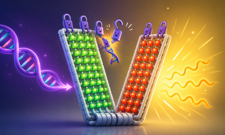
Featured image adapted from an AI image generator source and Selnihhin et al.
Every day, doctors and scientists look for tiny molecular clues hidden inside our bodies. This could be a fragment of viral DNA, a cancer-associated mutation, or a bacterial gene. Finding these molecules quickly can mean diagnosing disease earlier, choosing the right treatment, or stopping an outbreak before it spreads.
The challenge is that these targets are incredibly small. Detecting them often requires sophisticated laboratory equipment, specialised microscopes, or techniques like polymerase chain reaction (PCR) that multiply the amount of DNA before it can even be seen. That makes testing accurate, but not always simple or accessible.
So instead of building better microscopes, what if we built brighter biosensors?
That is exactly the question behind this study.
The researchers turned to one of the most unexpected building materials in modern nanotechnology: DNA. Most of us think of DNA as the molecule that stores genetic information. It carries the instructions that allow cells to grow, divide, and function. But over the past two decades, scientists have discovered that DNA has another remarkable talent. It can also be folded into precisely designed nanostructures, a technique known as DNA origami.
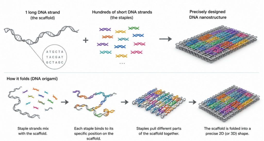
The idea is surprisingly simple. One long strand of DNA acts like a sheet of paper, while hundreds of much shorter DNA strands act like molecular staples. Each staple is designed to bind to a specific location, pulling different parts of the long strand together until it folds into a predetermined shape. By carefully designing these staple strands, researchers can build structures only a few tens of nanometres across, including boxes, tubes, hinges, gears, and even tiny robots.
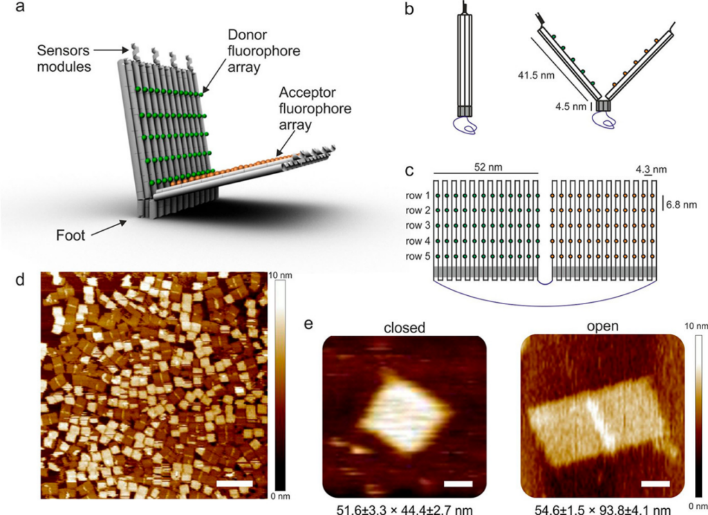
For this study, the researchers built something different. Instead of making another DNA nanostructure, they wanted to create a device that actually does something. The result was a tiny DNA origami beacon. At first glance, it resembles a miniature book standing upright. It consists of two rectangular DNA panels connected at the bottom by a rigid hinge. Along the top edge sit four short DNA “locks” that keep the two panels closed. As long as these locks remain intact, the beacon stays shut. But these locks are not ordinary pieces of DNA. Each one is designed to recognise a specific DNA sequence.
When the correct target DNA appears, it first binds to a short-exposed region called a toehold. This small interaction starts a process known as strand displacement, where the incoming DNA gradually replaces one of the strands making up the lock. Once enough locks are removed, the two DNA panels are no longer held together. Because DNA strands naturally repel each other due to their negatively charged backbones, the beacon swings open like a tiny book whose clasp has just been released. Opening the beacon is clever, but there is still one obvious problem.
How do you know it has opened? This is where the researchers introduced the real innovation.
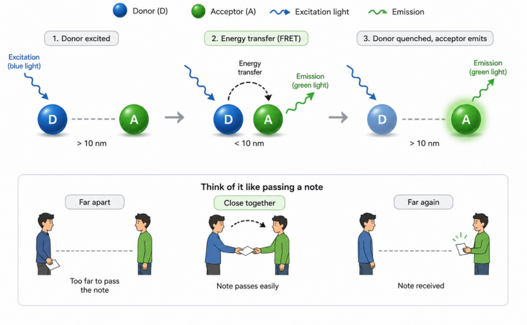
They decorated one panel with dozens of fluorescent donor dyes and the opposite panel with matching acceptor dyes. When the beacon was closed, the dyes sat close enough for energy to pass between them through a phenomenon called Förster Resonance Energy Transfer, or FRET. When the beacon opened, the dyes moved farther apart, reducing that energy transfer and changing the fluorescence signal.
If you have never heard of FRET before, think of it like two people passing a note across a room. When they stand close together, the note passes easily. Move them farther apart, and the exchange becomes much harder. Fluorescent dyes behave in a similar way. They only exchange energy efficiently when they are separated by just a few nanometers. Using FRET in biosensors is not new. The clever part of this work was making the signal much brighter.
Most DNA beacons rely on a single donor dye and a single acceptor dye. The researchers instead arranged entire arrays of fluorophores on the DNA origami structure, placing up to 60 donor dyes on one panel and 60 acceptor dyes on the other with nanometre precision. Rather than relying on one fluorescent pair, dozens worked together simultaneously. It is a little like replacing a single candle with an entire string of fairy lights. The principle stays the same, but the signal becomes far easier to see.
After testing different arrangements, the researchers found that using four rows of fluorophores, corresponding to 48 donor-acceptor pairs, gave the best balance between brightness and signal change. The beacon responded within about two minutes after the target DNA was added, and the stronger fluorescence allowed individual DNA origami devices to be detected using a conventional fluorescence microscope instead of specialised single-molecule instruments. Using this approach, they detected target DNA at concentrations as low as 100 picomolar.
What makes this design especially exciting is that the beacon itself does not need to be rebuilt for every new application. Only the DNA locks need to change. Replace those locks with sequences that recognise a different target, and the same DNA origami scaffold could, in principle, be adapted to detect other DNA molecules, RNA, proteins, or even small molecules associated with disease.
DNA is often introduced as the blueprint of life. This study reminds us that it can also become something entirely different. A scaffold. A hinge. A switch. A biosensor. Sometimes the same molecule that stores life’s information can also become the tool that helps us read it.

