Title: Multiple origins of green coloration in frogs mediated by a novel biliverdin-binding serpin
Authors: Carlos Taboada, Andrés E. Brunetti, Mariana L. Lyra, Robert R. Fitak, Ana Faigón Soverna, Santiago R. Ron, María G. Lagorio, Célio F. B. Haddad, Norberto P. Lopes, Sönke Johnsen, Julián Faivovich, Lucía B. Chemes, and Sara E. Bari.
Journal: PNAS
Year: 2020
If you have ever watched a frog sit on a leaf, you may have seen camouflage without realizing you were also observing chemistry. A frog’s green skin may seem like simple camouflage, but in some species, that color comes from an unexpected molecular trick: recycling a bile pigment.
The pigment is called biliverdin. If the name sounds familiar, it might be because biliverdin is known to cause the greenish color that appears as a bruise heals. As old red blood cells are recycled, the heme group inside hemoglobin is converted into biliverdin, a blue-green intermediate in the heme breakdown pathway. In many animals, biliverdin does not stick around for long; it is further processed or cleared away.
Some frogs, however, give biliverdin another path.
In several tree frogs and glass frogs, biliverdin is found at high concentrations in the blood, lymph, muscles, bones, and other tissues. This unusual accumulation of bile pigment is known as physiological chlorosis. Although elevated bile pigments can signal problems in other animals, these frogs have turned biliverdin into something surprisingly useful: a way to blend into their surroundings.
Still, this raises an important question: how do these frogs accumulate so much biliverdin, and how does it contribute to their coloration?
Taboada et al. found that the answer involves proteins called biliverdin-binding serpins, or BBSs. Serpins are a large family of proteins best known for inhibiting proteases, enzymes that cut other proteins. But in these frogs, certain serpins have taken on a different role. By binding biliverdin, these proteins alter the pigment’s light-absorbing properties, helping turn a product of heme breakdown into biological color.
This is where the chemistry comes in. A pigment’s color does not depend only on the pigment itself. It also depends on the pigment’s molecular environment.
Think about how different the same object can look under sunlight, fluorescent lights, or the warm glow of golden hour. The object has not changed, but the lighting affects what you see. Molecules behave a little differently: their absorption of light depends on their electronic structure, which can be influenced by their shape and interactions with surrounding molecules.
In the case of BBSs, biliverdin binds noncovalently to the serpin protein. The researchers found that each protein carries approximately one biliverdin molecule. Although the study did not determine the precise atomic structure of the binding site, spectroscopic measurements suggested that biliverdin adopts a partially extended conformation within the protein. Rather than behaving like a free pigment in solution, biliverdin exists in a protein environment that changes the way it absorbs light.
Those differences can be measured. When a molecule absorbs light, its electrons undergo transitions between energy states. Changes in molecular conformation and environment can alter the energies and intensities of these transitions, changing the wavelengths and amounts of light absorbed.
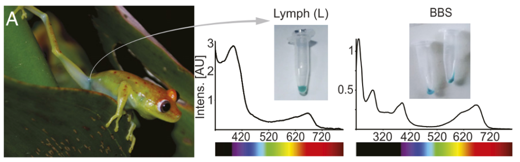
The researchers observed this effect directly by isolating BBS from the blue-green lymph of the tree frog Boana punctata. After removing yellow pigments and other components, they obtained a purified protein that appeared cyan. Its absorption spectrum showed two prominent peaks, one near 390 nanometers and another near 667 nanometers. Compared with free biliverdin, the relative intensities of these peaks were markedly different, providing evidence that the protein-bound pigment adopts a different molecular conformation.
Figure 1 illustrates this connection between molecular chemistry and visible color. The blue-green lymph can be seen beneath the frog’s translucent skin, while the isolated BBS retains a striking cyan appearance. The absorption spectra reveal that this color is not simply a property of biliverdin alone: its interaction with the serpin changes how strongly it absorbs different wavelengths of light.
But the frog’s final green appearance is not produced by biliverdin alone. Instead, it results from a layered optical system involving cyan BBS, yellow pigments, and structures within the tissues that reflect and scatter light. In B. punctata, light travels through the translucent skin and underlying lymph before interacting with deeper tissues, including a reflective layer containing guanine crystals.
The authors used an optical model to investigate how these layers work together. They found that BBS plays an important role in reducing the amount of red light reflected by the frog, particularly around the wavelengths absorbed by protein-bound biliverdin. This helps produce a sharp increase in reflectance near 700 nanometers, similar to the characteristic red edge seen in green vegetation. As a result, the frog’s reflectance more closely resembles that of the leaves around it, even extending into the near-infrared region of the spectrum. In other words, BBS does more than make the frog look green: it helps the frog blend into its environment across wavelengths beyond those visible to humans.
Even more remarkably, this strategy appears to have evolved more than once. The researchers identified physiological chlorosis in more than 430 frog species and inferred at least 41 independent evolutionary origins. By studying BBSs from multiple frog lineages, they found that different serpins had evolved the ability to bind biliverdin, suggesting that evolution arrived at similar molecular solutions on multiple occasions.
For chemists, that is the exciting part. This system is not just a fun frog fact. It shows how living organisms can tune the behavior of small molecules by changing their molecular surroundings. Biliverdin is usually associated with heme breakdown. Serpins are usually associated with protease regulation. But through evolution, a familiar pigment and a familiar protein family have become tools for camouflage.
So what does it take to make a frog green?
First, recycle a bile pigment.
Then, bind it to a specially evolved protein.
Finally, use light itself to blend into the leaves!