
Title: Nucleated synthetic cells with genetically driven intercompartment communication
Authors: Ion A. Ioannou, Carolina Monck, Francesca Ceroni, Nicholas J. Brooks, Marina K. Kuimova, and Yuval Elani
Journal: PNAS
Year of publication: 2024
Doi: https://doi.org/10.1073/pnas.2404790121
The creation of synthetic cells involves designing and engineering systems which replicate a biological cell’s function and structure. The importance of this? It provides a model system to study biological processes, understand cellular functions and cellular signaling, as tools for drug delivery, and to provide clues about how life started.
The quest to create a synthetic cell akin to a eukaryotic cell with compartments that mimic the cytoplasm, nucleus and other organelles while also capturing the functionalization of these components has been challenging.
Ioannou et al have developed a method to successfully create a cell-sized assembly with a nuclear compartment and a cytoplasmic compartment. They have also genetically engineered communication between these compartments. This replicates key features of a biological cell where communication between cellular compartments and subsequent biomolecular reactions are observed.
To design this vesicle-in-vesicle cell, they first created a column containing 4 liquids with different densities. The first layer had mineral oil (o1), the second had glucose water (w1), the third had silicone oil (o2) and the fourth contained sucrose in water (w2). In each of the oil layers, the scientists incorporated lipids with a poly-ethylene glycol molecule attached to it (PEGylated lipids). The different densities of the liquids led to the formation of 4 liquid layers with a lipid-water interface between each layer. Next, a droplet of water-oil emulation containing maltotriose (w3) was deposited at the top of the column. This droplet had the highest density, and it travelled through the column as the assembly was centrifuged. (Fig 1 A)
As the drop passed through each interface, a lipid monolayer was deposited on the droplet. Thus, at the bottom of the column, the emulsion droplet formed the nucleus compartment, the glucose water formed the cytoplasmic compartment and the environment of this “cell” was the sucrose water. The lipid layers formed the membranes for these compartments and incorporation of PEGylated lipids allowed them to increase the success rate of cell formation and prevented the membrane layers from interacting with each other. (Fig 1A)
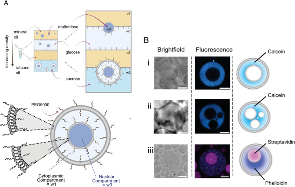
They confirmed the compartmentalization and generation of nucleated cells by incorporating a dye called calcein in the glucose water. Under microscopy, 74% of the successfully generated cells showed a ring-shaped structure indicating the formation of a distinct “cytoplasmic” compartment (Fig 1B i). Later, they incorporated complex biomolecules in it. They loaded a molecule called alexa647-phalloidin in the cytoplasmic compartment and alexa488-streptavidin in the nucleus. Using microscopy, they could visualize both the biomolecules (Fig 1B ii). This suggested that these systems could be loaded with complex macromolecules, thereby replicating the multi-component nature of a cell.
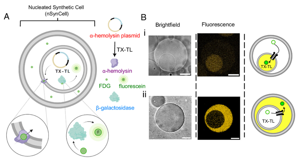
One necessary aspect of synthetically modeling a biological cell is replicating its its functionality. A major advantage of creating a nucleus was being able to transfer material from the nucleus to the cytoplasmic region, as happens in the biological cells where RNA and other proteins shuttle to and from the compartments. To replicate this functionality, they chose to express a membrane pore protein called α-hemolysin which transports a chemical signal between the two compartments. The expression of this protein was achieved by using a system called transcription and translation (TX-TL) which allows for gene expression outside of cellular systems (in vitro).
Fig 2A shows the schematic for this genetically engineered cell. In this model, a non-fluorescent substrate called FDG is encapsulated in the cytoplasmic component. The TX-TL system containing beta-galactosidase (β-gal) and the α-hemolysin DNA will be encapsulated in the nucleus. When the α-hemolysin is expressed and incorporated into the inner membrane, the FDG will flow from the cytoplasm into the nuclear compartment where the β-gal will degrade it to a fluorescent molecule called fluorescein. Under fluorescent microscopy, they observed fluorescence in the nuclear compartment of those cells where α-hemolysin was expressed (Fig 2B i). Furthermore, they reversed the order by loading the FDG in the nucleus and the TX-TL-β-gal along with α-hemolysis in the cytoplasm. They noticed that FDG, being smaller than the pore size, moves from the nucleus into the cytoplasm, combines with the β-gal and produces fluorescence (Fig 2B ii). They also observed that the size, chemical complexity and osmolarity of the encapsulated solutions directly affected the yield and the diameters of the cells.
The authors successfully developed an accessible and easy method to create a nucleated synthetic cell which mimics a eukaryotic cell by allowing genetic modifications, protein expression, and intercompartment signaling. This work addresses the current limitations with synthetic cells by gaining control over the composition of the compartments, and paving way to develop functionally complex cells and expanding our understanding of the compartmentalization in biological cells.