
Article: Development of a Hydrogel Platform with GBM and Microglia: A Potential Glioblastoma Tumor Model
Authors: Seyma Isik, Deniz Yucel, Vasif Hasirci
In: ACS Applied Bio Materials
The brain is largely made up of cells that reside in a space called the extra-cellular matrix (ECM). This is typically made up of hyaluronic acid, glycosaminoglycans and proteoglycans which keep the ECM soft and hydrated. When cancer (glioblastoma, a brain cancer, in this case) strikes, the ECM undergoes changes in its composition, and it becomes stiffer. Studying the ECM, and the interaction of different cells with it becomes an integral part of understanding cancer pathology, since cancer is a disease which spreads in the body through cell migration and invasion. However, replicating the ECM in a lab environment has been a challenge for a few years.
The most recent and biologically closest ECM model is the decellularized tissue, which refers to a tissue from which all the cells are removed, leaving only the ECM behind. But this suffers from multiple limitations, including the effectiveness of decellularization and altered composition of proteins. Isik et al have developed a model which replicates the glioblastoma ECM in a biological and mechanically accurate manner, by combining decellularized tissue with hyaluronic acid methacrylate (HAMA).
To develop this model, they first decellularized brain tissue from cows, and assessed the content of a range of ECM components. They optimized the process until the decellularized tissue mimicked the native ECM in the brain. Once this was ready, they solubilized it in hydrogels. A hydrogel is a polymer that can absorb and retain large amounts of water. In this case, it was made of methacrylate hyaluronic acid (HAMA), and the decellularized tissue powder was dissolved in it. It was then allowed to undergo a process called gelation (where a liquid transforms into a gel, creating a 3-D network). For this, they exposed the solution of HAMA and decellularized tissue to ultraviolet radiation for 2 minutes and allowed cross linking and swelling for 24 hours. At the end, they had created a hydrogel-decellularized tissue complex, called 1H3D, which had similar properties to glioblastoma tissue, such as a similar water content, compression moduli, and elasticity. Moreover, this complex degraded much more slowly, and had a superior mechanical strength, making it an ideal model system to study glioblastoma.
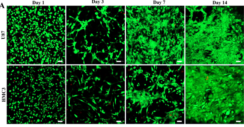
They next wanted to study the behavior of cells within the 1H3D matrix. They entrapped glioblastoma cells (U87) and microglial cells (HMC3; immune cells of the brain) in it, and found that they were viable over 7 days. The cells formed cellular networks (branched structures and elongations) within the hydrogel, and their density increased over time (Fig. 1).
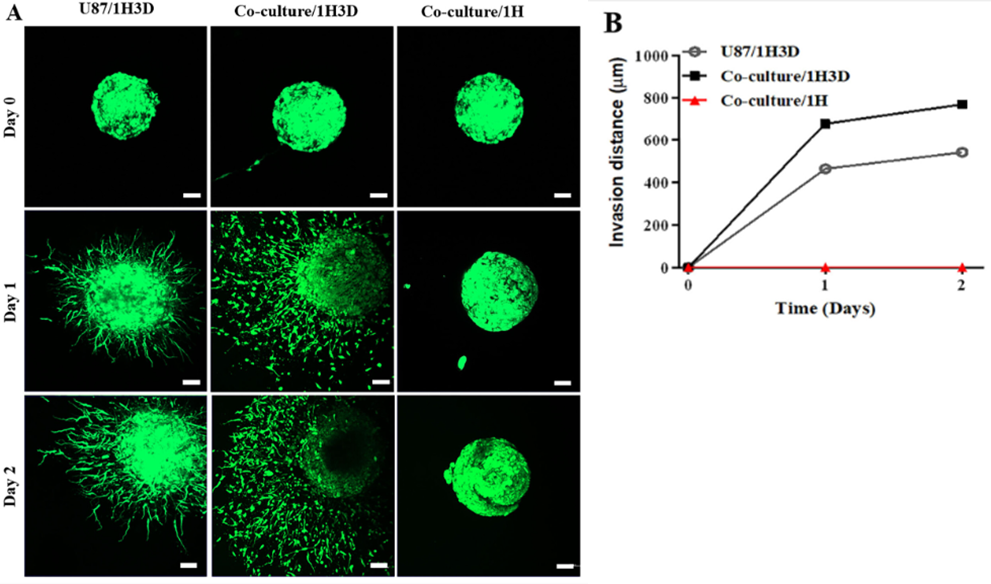
An important characteristic of glioblastoma is that it is invasive, i.e., it infiltrates healthy brain tissue. The authors studied this property of glioblastoma in the 1H3D hydrogel and the HAMA hydrogel. They found that 1H3D hydrogel provided the perfect environment in which the U87 cells showed increased invasion and cell migration. Further, the invasion of U87 cells was further increased when they were co-cultured it with HMC3 cells in the 1H3D hydrogels, suggesting the important role of microglial cells in the tumor microenvironment (Fig. 2).
Overall, the authors have created a model system that closely resembles the glioblastoma ECM composition and its behavior in this ECM. A limitation of the study is that it doesn’t test with patient-derived tumor cells, incorporate the multiple cell types and the tumor microenvironment found in the brain. These are interesting avenues that can be explored in the future. While the study has these limitations, it paves way for future studies to understand tumor progression and novel cell signaling pathways.
Photo by National Cancer Institute on Unsplash.