
The physiological level of molecules like ATP (Adenosine Triphosphate), can indicate brain health and neurodegenerative diseases such as Alzheimer’s. As we age, the levels of these neurotransmitters in our brain – like ATP, can run low and could be a sign of possible health risks.
Ever wondered if we could peek inside the brain to spot early signs of such disorders? Turns out, it is not that straightforward, thanks to the protective blood-brain barrier (BBB). The BBB regulates and prevents the uptake of any non-essential molecules by the brain. Getting any sensor through this barrier is a persistent challenge being addressed by several researchers globally.
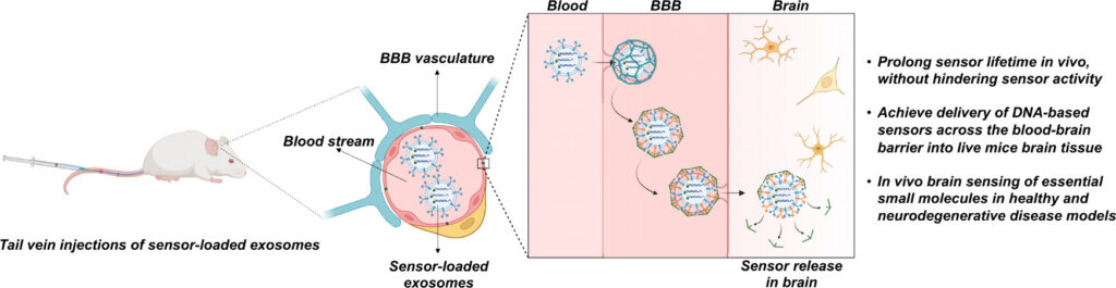
The Lu Lab at the University of Texas at Austin have figured out how to use brain-cell-derived exosomes to deliver sensors across the BBB. You could think of exosomes as tiny messengers, carrying the sensors and sneaking past the heavily guarded barrier to the brain. These exosomes derived from brain cells, are perfectly disguised as they are rich in proteins and other macromolecules that are inherently recognized by the BBB. This makes exosomes biocompatible, specific and have a higher delivery efficiency. The sensors they use is an ATP-responsive aptamer, which is a single-stranded DNA molecule that can bind to ATP! This development has allowed for improved brain imaging in mouse models of Alzheimer’s disease.
In lab models and mouse studies, the exosome-packed sensors crossed the BBB much more efficiently than traditional delivery systems. In an Alzheimer-mouse model, the researchers could pinpoint areas like the hippocampus and cortex, where ATP levels were unusually low and could be classic Alzheimer’s hotspots
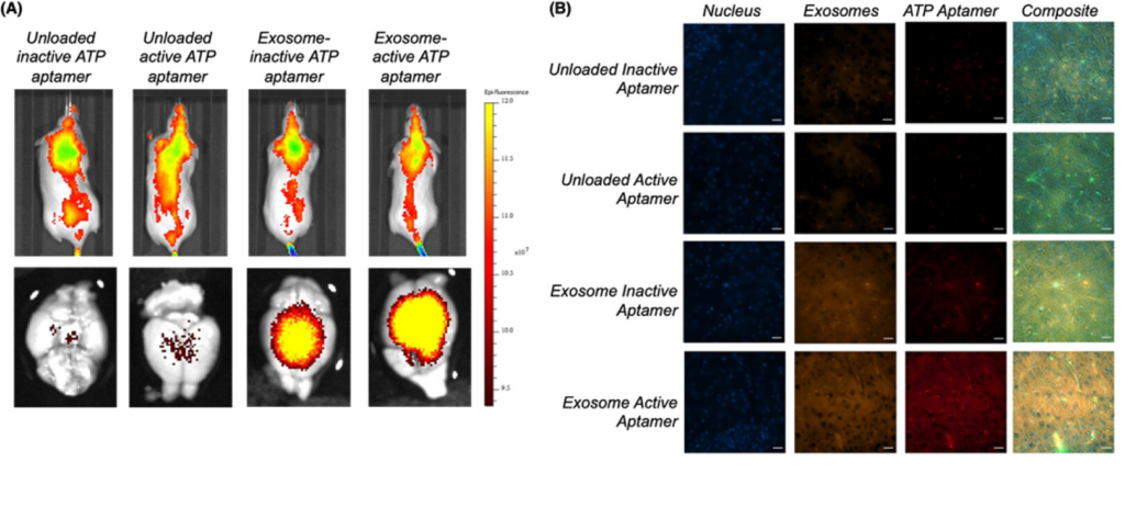
“While previous Alzheimer’s research studies have also found decreased levels of both targets in whole brain tissue and patient serum, we were able to provide spatial resolution in live tissue, demonstrating that the decreased levels of ATP were not uniform throughout the brain,” they stated.
The best part? This delivery system could be adapted for all sorts of molecules. All you would need to do is to have an aptamer selective for the metabolite of your choice and use a similar delivery strategy to study how it is modulated. This could significantly improve the future of brain health diagnostics!