

| Title | Improvement in protein HSQC spectra from addition of betaine |
| Authors | Finn O’Dea, Aiden J. Seargeant, Jessica Hurcum, Rodolpho do Aido-Machado, Nicola J. Baxter, Jon P. Waltho, Jon R. Sayers, Mike P. Williamson |
| Journal | Journal of Biomolecular NMR |
| Year | 2025 |
| URL | https://link.springer.com/article/10.1007/s10858-025-00463-0 |
| Open Access? | Yes! No paywall |
When you look at the pharmaceutical offerings on the current market, the most impactful treatments often involve biologics, a shorthand term for biological compounds. These products can be proteins, like the monoclonal antibody Datroway used to treat breast cancer, or even complex mixtures, such as cellular Lantindra used for Type 1 diabetic patients with severe hypoglycemia complications. However, unlike other small molecule drugs like aspirin, biologics have greater storage concerns and a shorter shelf life before their efficacy runs out. For example, Datroway is shelf stable for 3 years when unopened and dry – but once reconstituted in water, the antibody must be administered within 24 hours. In contrast, aspirin remains 90% effective even after five years when stored at room temperature after opening the bottle.
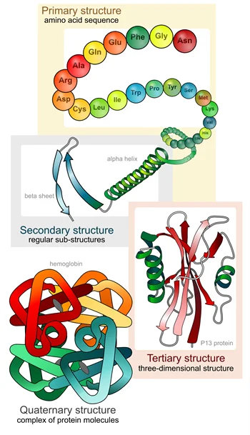
The shorter stability of biologics is likely due to an undesired change in protein structure. More than 60% of our proteins are folded in a specific way every time they are made (Fig 1), and even a small change in the structure could prevent their intended biological function, necessitating sensitive quality control checks before a product is made available to a patient. Pharmaceutical companies ensure good quality products by following extensive quality control regulations enforced by governmental agencies. However, tracking down minor changes in a protein’s structure is difficult due to the low sensitivity of current instruments and methods. Recently, scientists figured out a new way to enhance the detection of protein structure at atomic-level resolution with the addition of a single molecule.
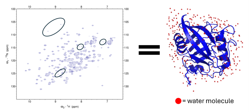
Pharmaceutical companies like Sanofi and Merck already used nuclear magnetic resonance (NMR) to study their small molecule drugs. They have expanded their use of the technique to study the individual small molecules, called amino acids, that make up the proteins. This powerful biophysical method allows them to detect if even one amino acid is out of alignment from its normal structure. Scientists normally accomplish this by obtaining an HSQC spectrum of a protein, which contains one “peak” per amino acid corresponding to the hydrogen-nitrogen bond (with a few exceptions). The HSQC spectrum can then become the “fingerprint” that identifies the protein every time it is made, as the position of each peak on the spectrum represents the amino acid type and its specific position within the protein structure (Figure 2).
However, some parts of the protein are more dynamic. This can lead to some amino acids in the protein becoming invisible on NMR spectra, much like the Flash does when he runs at his superspeed! This is due to the exchange rate of the hydrogens attached to the protein (called amide hydrogens) with the hydrogens attached to the water molecules in the solution. The exchange rate for the protein hydrogens with water hydrogens can be up to 1000 times faster (a timescale of microseconds to nanoseconds) than the rate of the NMR measurement (a timescale of microseconds to milliseconds), and lead to their NMR peaks to be small and weak in comparison to more static protein hydrogens.
Scientists in the Williamson group at the University of Sheffield, United Kingdom, figured out that by adding betaine, a compound extracted from sugar beets, the exchange rate of the protein hydrogens with water decreased by a third. And like plunging the Flash in a vat of molasses, the increased viscosity helped the scientists enhance the signal of these weaker peaks and better see them.
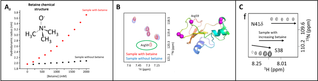
They showed the usefulness of their method by adding betaine to a protein called Barnase, which undergoes small changes in structure as it degrades RNA. If something is wrong with Barnase and the desired reaction doesn’t proceed, it could be difficult to determine why if the method isn’t sensitive enough to small changes in protein structure. They figured out that betaine molecules provided a protective coating around the protein, enlarging its hydrodynamic radius and pushing away the water molecules that normally exist close to the protein surface (Fig 3A). This did not change the structure or solubility of Barnase, but did enhance weak peaks in its HSQC spectrum. One of these peaks corresponded to an amino acid involved in the chemical reaction called Arg59. The scientists saw that Arg59 appeared only in the betaine-containing solution (Fig3B). Another residue, Ser38, showed a significant enhancement of signal with increasing amounts of betaine, further demonstrating the signal enhancement possible (Fig 3C).
One major downside to the method is that it is not specific in decreasing the amide exchange rate of only the fast protein hydrogens – it also hits the slow exchanging hydrogens. This resulted in about a 25% average signal reduction across all protein hydrogens. However, for many proteins, the pros of seeing weak peaks can outweigh the cons of global signal reduction. Once the method has been validated with a wide array of proteins, this method has great potential to become standard practice for industry quality control to enhance the structure “fingerprint” for a variety of biologics. This could vastly improve our quality control methods at the final step, ultimately benefiting patients and ensuring only functional proteins are waiting on the pharmacy shelves for use.