
Authors ~ Saugat Mondal, Jusung An, Tapas Bera, Moumita Banerjee, Snehasish Debnath, Debasish Mandal, Antara Sikder, Samit Guha, Jong Seung Kim, N. D. Pradeep Singh
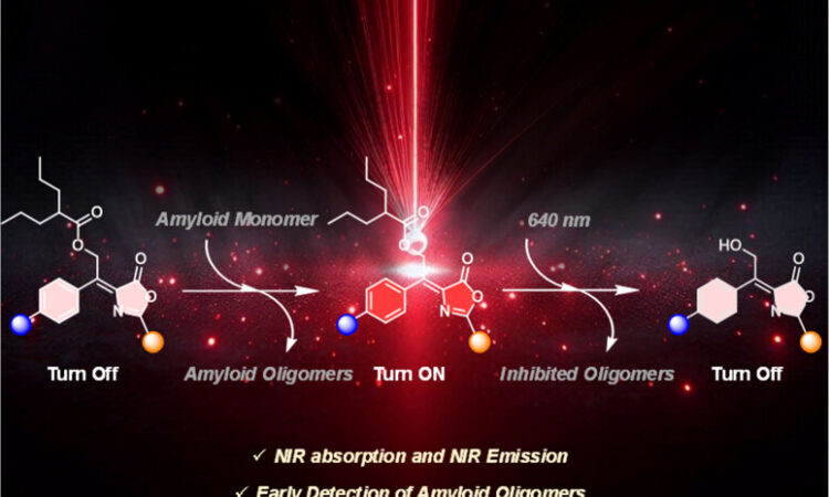
Scientists have developed a new strategy to release medicine in the brain using near-infrared (NIR) light, a wavelength known for its ability to penetrate biological tissues with minimal damage. At the center of this approach is a specially engineered “photocage,” designed from the green fluorescent protein (GFP) that gives jellyfish their natural glow. By modifying the GFP core, the researchers created a system that can both detect harmful protein clumps linked to Alzheimer’s disease and release therapeutic molecules when activated by light.
A “photocage” is a clever chemical wrapper that keeps a drug locked up until light tells it to open. Most older versions only worked with harmful ultraviolet light, limiting their medical use. This team flipped the script by designing photocages that respond to near-infrared (NIR) light—the same kind used safely in medical imaging. NIR light penetrates tissues deeply without damage, making it an ideal “key” to unlock medicine in precise spots inside the body. The researchers built their light-sensitive cages using the chemical skeleton of GFP’s glowing core. By tweaking this structure, they created a family of molecules—called meso-GFP-PRPGs—that can both tag proteins and release tiny drug molecules when illuminated.
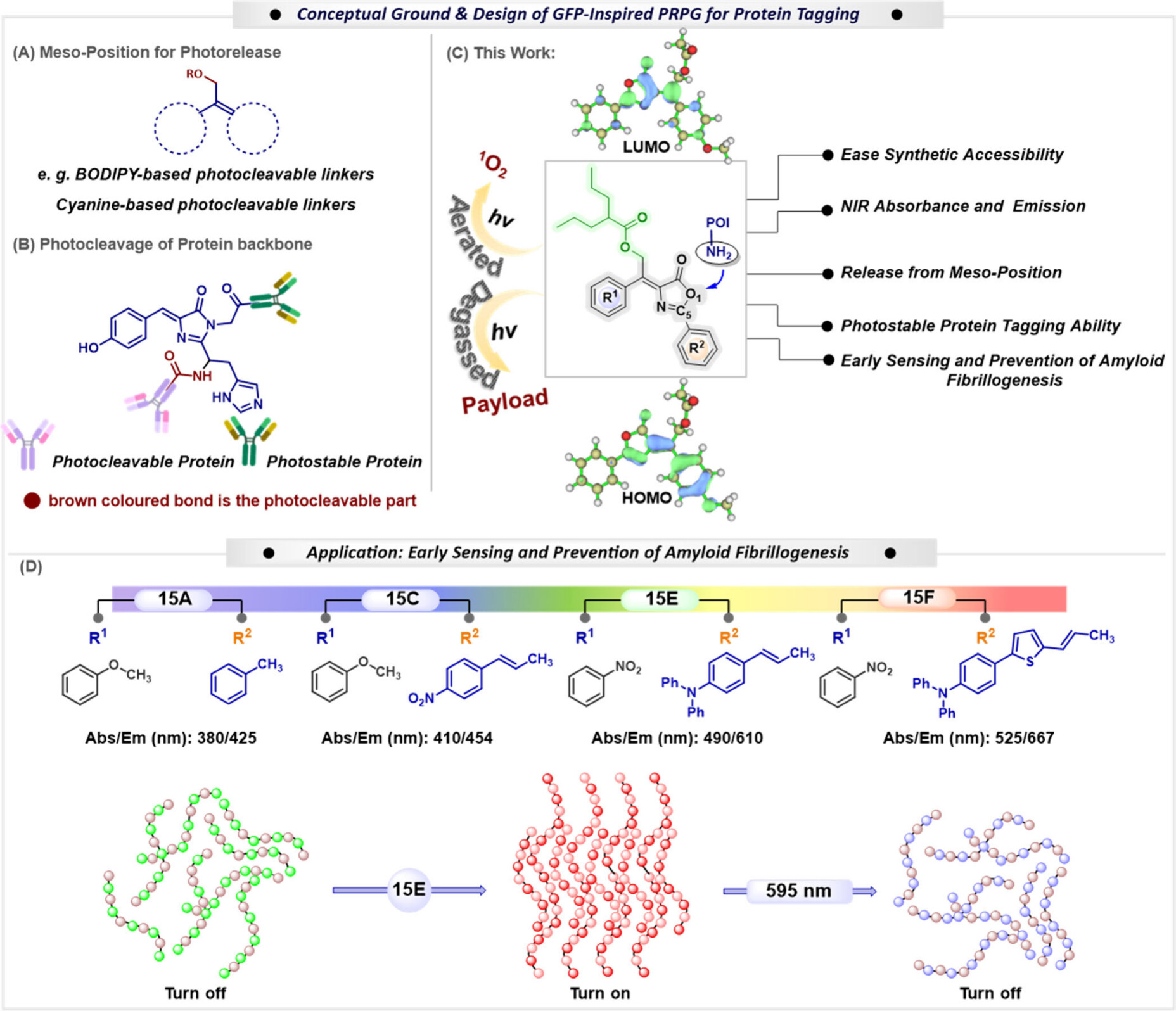
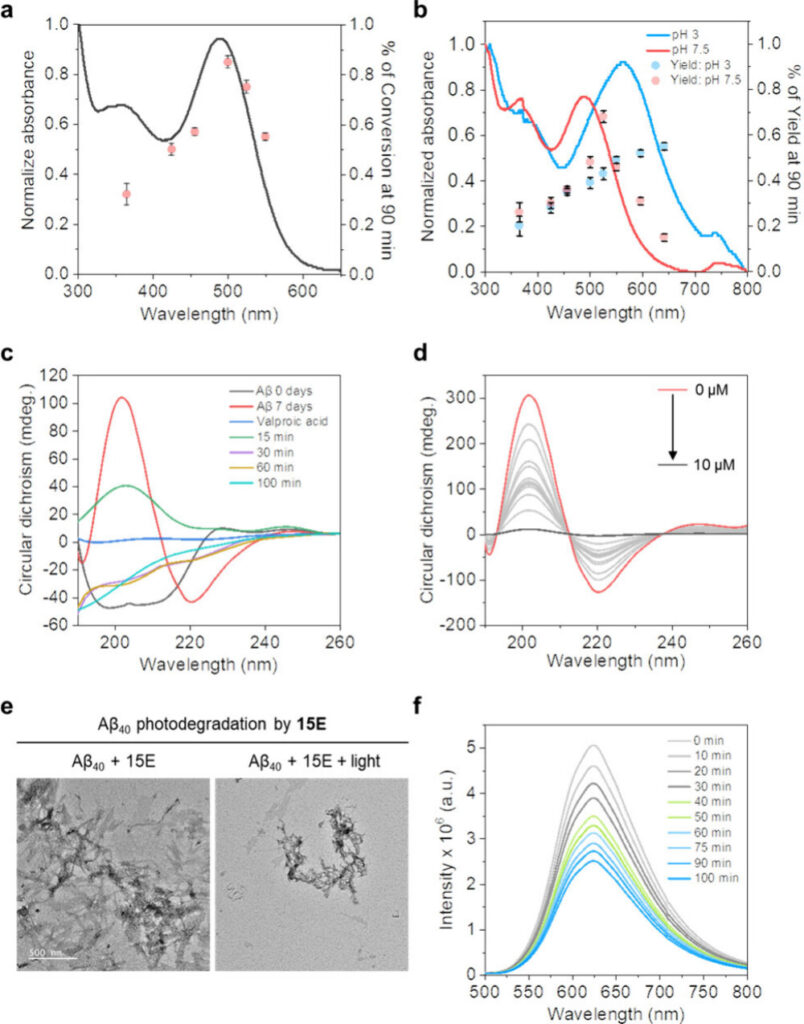
Alzheimer’s disease is characterized by the accumulation of amyloid-beta protein clumps in the brain. One molecule in particular, 15E, showed exceptional performance: it could detect amyloid-beta oligomers and fibrils with much higher sensitivity than conventional dyes, and when activated by red light, it released valproic acid, a neuroprotective drug. The released drug helped break down harmful amyloid fibrils, showing both diagnostic and therapeutic potential. Laboratory experiments confirmed that 15E not only highlighted amyloid aggregates with strong fluorescent signals but also reduced their toxic effects on nerve cells when combined with light. This dual functionality—detecting disease-related proteins and delivering treatment on demand—demonstrates the promise of these photocages as theranostic tools.
While more research is needed before clinical applications, this work illustrates how re-engineering the chemistry of a natural fluorescent protein can lead to innovative strategies for tackling neurodegenerative diseases. By merging biomolecular inspiration with light-activated precision, this can serve as a new blueprint for next-generation drug delivery systems.