
| Title | Nanoparticles from Cuttlefish Ink Inhibit Tumor Growth by Synergizing Immunotherapy and Photothermal Therapy |
| Authors | Rong-Hui Deng, Mei-Zhen Zou, Diwei Zheng, Si-Yuan Peng, Wenlong Liu, Xue-Feng Bai, Han-Shi Chen, Yunxia Sun, Pang-Hu Zhou, Xian-Zheng Zhang |
| Journal | ACS Nano |
| Year | 2019 |
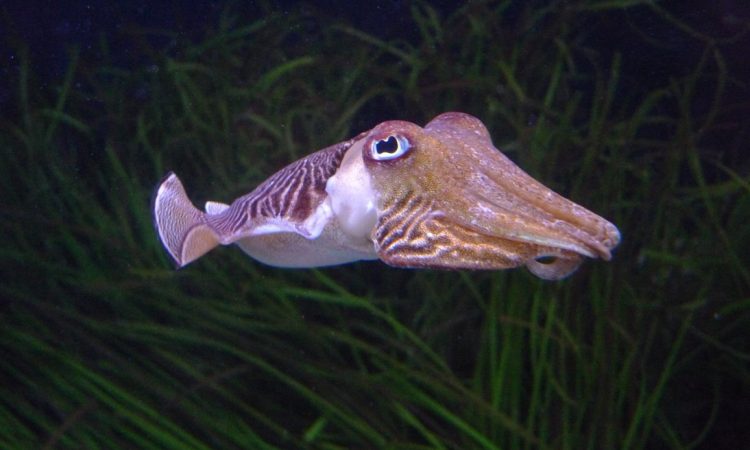
The more we learn about cephalopods the more we’re amazed by their sophisticated biology, but their brains may not be the only thing of interest to humans. A recent study describes nanoparticles from cuttlefish ink as a promising new cancer treatment (in mice).
We often think of nanoparticles as objects made by scientists, usually of metals like gold or silver – but nanoparticles are also found in nature. Cuttlefish ink, for instance, is made of nanoparticles, or particles less than 100 nm in size, which are made of a combination of melanin (which give the ink its dark color), polysaccharides (or sugars), oligopeptides (or short proteins), and metals. Interestingly, some of these polysaccharides may be able to stop tumor growth.
The nanoparticles target cells called tumor-associated macrophages – white blood cells that have been hijacked to aid tumor growth. The polysaccharides in the nanoparticles can “reprogram” the cells to behave like healthy cells, which would significantly slow the cancer progression.
Additionally, the high amount of melanin in the nanoparticles easily absorbs near-infrared light, making it a perfect target for photothermal therapy, an alternative to radiation therapy where incoming light is directed at a target, which absorbs and then releases that energy to kill the surrounding cancer cells. Melanin is a natural alternative to many of current targets used for photothermal energy, usually metal nanoparticles.
Researchers at Wuhan University in China characterized cuttlefish ink nanoparticles and found them to be not only biocompatible, but effective in reprogramming cancer cells in culture.
In determining whether cuttlefish ink nanoparticles are useful for cancer therapy, perhaps the most important factor is whether they are safe when injected into the bloodstream. When the researchers injected the nanoparticles into mice, they found that over time the ink did not change the biochemistry of the liver and kidneys (Figure 1). They also injected an excess of ink into a cell culture (more than would ever be used for therapeutics) and less than 5 percent of the cells died.
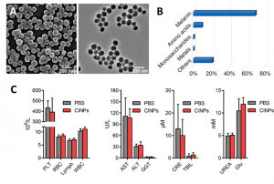
Figure 1. (A) Scanning electron microscopy images of cuttlefish ink nanoparticles. (B) Chemical composition of cuttlefish ink nanoparticles. (C) Concentration of certain biomarkers in blood, liver, and kidney with and without nanoparticle injection. Note that the levels do not change, showing that the nanoparticles are biocompatible. Adapted with permission from Deng, R. H., et. al. ACS Nano 2019. Copyright 2019 American Chemical Society.
- ACS Nano 2019. Copyright 2019 American Chemical Society.
While the ink was clearly safe for healthy organs in the mice, tumors did not fare so well against the nanoparticles. When cultured cancer cells were incubated with the nanoparticles, biochemical analysis showed that they were chemically “reprogrammed” to a healthy state. While this treatment alone wouldn’t kill a tumor, it would slow growth significantly by removing a mechanism for tumor growth.
The researchers then tested the cuttlefish ink nanoparticles as an agent for photothermal therapy. Photothermal therapy only works when the cells are irradiated with the correct wavelength of light – in the case of melanin this is in the near-infrared range. When the ink nanoparticles alone were introduced to tumor cells, they were not cytotoxic, or deadly. Only when radiated with 808 nm (near-infrared) light did tumor cells start to die. After radiation of cells with a 200 µg/mL concentration of nanoparticles, 90 percent of tumor cells died. A similar experiment in mice showed that after 6 minutes of irradiating a tumor, the temperature increased to 50 °C, which is enough to kill tumors. Additionally, the nanoparticles were shown to halt metastasis, or the spread of cancer to other organs, in another mice model.
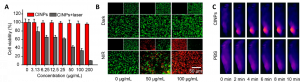
Figure 2. (A) Cell viability when treated with cuttlefish in nanoparticles. The nanoparticles alone do not kill cells (red), but when radiated with near-infrared light, the cells die (gray). (B) Fluorescence image of cells treated with cuttlefish ink nanoparticles. Green indicates living cells and red shows dead cells. Cells clearly died when 100 µg/mL of nanoparticles in cells were radiated with light. (C). Heat images of rats showing that tumors increased in temperature when nanoparticle-treated tumors were irradiated with near-infrared light. The effect was not nearly as great when a laser was directed at a tumor without nanoparticles. Adapted with permission from Deng, R. H., et. al. ACS Nano 2019. Copyright 2019 American Chemical Society.
Over and again, the key to some of the most effective medical interventions have existed in nature all along, from aspirin to penicillin. Cuttlefish ink is a promising treatment not only for its efficacy or “natural” credentials, but because a biological source for new cancer treatments subverts the need for complicated and costly synthetic strategies. Hopefully in the coming age of our cephalopod overlords, they’ll mercifully donate their ink.
Feature image courtesy of Wikimedia Commons.