Title: Two Water Two Water-Soluble and Wash-Free Fluorogenic Probes for Specific Lighting Up Cancer Cell Membranes and Tumors
Authors: Shumin Feng, Yijia Liu, Qianhua Li, Zhisheng Gui, and Guoqiang Feng
Journal: Analytical Chemistry
Year: 2022
DOI: https://doi.org/10.1021/acs.analchem.1c03685
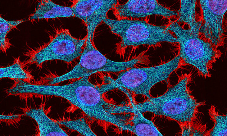
Featured image by Tom Deerinck and NIH is licensed under CC BY-NC 2.0
There are numerous analytical methods for the study of cancer and tumors. Cancer cells have distinct features that differentiate them from healthy cells. Researchers take advantage of these unique properties to detect, analyze and follow the progression of cancer cell development. An interesting cellular structure for cancer research involves the cell membrane, which is a thin membrane that encloses a cell. The cell membrane serves two general functions; to act as a barrier to protect the cell and to act as a gate for the exchange of materials (e.g. nutrients) in and out of the cell. The cell membrane plays a crucial role in stability of the cell and therefore is important biological structures of study.
Cell membranes are too thin to be clearly observed using a light microscope, so alternative detection methods are required. Selective visualization methods are important because they allow for biomedical diagnosis and research allowing researchers to detect and follow the cells during their progression of cancer development.
An analytical method for studying cell membranes include fluorescence analysis with use of a fluorogenic probe to visualize the cell membrane. Current probes include membrane-associated probes and polarity sensitive probes. Membrane based probes support study of the cell membrane while polarity probes are capable of monitoring changes in cellular polarity. Polarity is an important environmental factor for a cellular functions as it impacts various physiological and chemical processes such as protein activation, enzyme activity, signal transduction processes and membrane systems. This is significant as abnormal changes in polarity contribute to development of diseases such as diabetes and Alzheimer’s.
In a study by Feng et al., (2022) the authors developed two novel fluorogenic probes which are collectively called MEMs. These fluorogenic probes are unique as they incorporate properties of both membrane associated and polarity sensitive probes. Figure 1 shows the structure of the MEMs probe which consist of a lipophilic group (dissolve in fats/lipids) and hydrophilic group (dissolve in water). The MEMs probe is further categorized as two specific probes known as MOM and MOS where they have different functional “R” group. These fluorogenic probes have capacity to selectively target cancer cell membranes where it emits strong red fluorescence light in low polarity conditions.

The MEMs probe demonstrate that cancer cells potentially have lower cell membrane polarity than normal cells, which allow for an effective fluorescence method for cancer cell detection and analysis. Feng et al., (2022) further demonstrate this through a cancer cell imaging study with MEMs (figure 2). In the study, they applied the MEMs probe to various cancer cells, called HepG2, HeLa, A549 and U87-MG, and normal cells, called RAW264.7, LO2, and HEK 293T. Each cancer cells represent a different type of cancer. For instance, HepG2 is for hepatocellular carcinoma (human liver cancer), Hela for cervical cancer, A549 for human lung carcinoma and U87-MG for malignant gliomas (brain tumor). In contrast, the normal cell RAW264.7 is a cell model with macrophage-like properties to help model immune system, LO2 to model human liver and HEK 293T for embryonic kidney cells.

The study in figure 2a observes MOM with bright field and fluorescence (red channel) microscopy. The merge is the overlap between bright field and red channel. The strong red fluorescence from MOM is only observed for cancer cells under the red channel. The strong selectivity for cancer cells is further evident in Figure 2a and c, which demonstrates that MOM and MOS display significantly stronger signal (greater red fluorescence intensity) for cancer cells than normal cells. The study highlights that MEMs are capable of discriminating cancer cells from normal cells, providing an effective fluorescence method for cancer study. In addition to strong detection, these fluorogenic probes are also advantageous as they are water soluble and wash free, meaning no additional processes are required to separate stained cells from unbound fluorescent probes Furthermore, the probes exhibit low toxicity towards cells, and are photostable, allowing for development of clear and visible fluorescent images.
Feng et al. developed two novel fluorogenic probes that are collectively referred to as MEMs (specifically MOM and MOS), which are capable of effectively distinguishing cancer cells from normal cells based on polarity differences. The development of the novel fluorogenic probes provide an efficient and simple method for cancer cell detection with significance for cancer research and diagnosis.

